MagViT, an interpretable multi-magnification transformer framework with scale-gated fusion and patient-level model selection, is presented, an interpretable multi-magnification transformer framework with scale-gated fusion and patient-level model selection relative to prior ViT-centered BreakHis work.
Abstract
Breast cancer is one of the most common types of cancer among women around the world. Rapid detection and early treatment can hinder its progress to more complex stages and can impede its spread to other parts of the body. Histopathological image classification is the most common task in cancer detection due to its robustness in analyzing cellular data. Breast histopathology classification requires handling both multi-scale tissue morphology and clinically relevant generalization beyond the source domain. This paper presents MagViT, an interpretable multi-magnification transformer framework with scale-gated fusion and patient-level model selection. The model uses four BreakHis magnifications (40X, 100X, 200X, 400X) and extracts per-scale representations with a ViT backbone, and combines them via a learnable gate that masks missing scales. Patient-level five-fold cross-validation with a fixed seed has been run and compared with three architectural branches. The most accurate branch is then selected as the final model due to the strongest patient-level accuracy while retaining the simplest fusion pathway. On BreakHis, our architecture achieves a mean image accuracy of 0.9191, a mean patient accuracy of 0.9643, and a mean macro-F1 of 0.9042. External transfer experiments provide preliminary evidence of cross-dataset generalization under controlled adaptation settings on BUSI (image accuracy 0.8306, macro-F1 0.7480, patient accuracy 0.8291) and IDC (image accuracy 0.8577, macro-F1 0.8191, patient accuracy 0.8372). Grad-CAM visualization indicates that the model focuses on diagnostically significant and meaningful regions across magnifications. Relative to prior ViT-centered BreakHis work, this study emphasizes patient-level selection and cross-dataset robustness under a reproducible protocol.
The integration of these advanced techniques significantly enhances diagnostic reliability, addressing the challenges in histopathological image analysis, and proposes a novel explainable multi-model DL framework for breast cancer classification leveraging histopathological images.
Muhammad Nabeel Mehmood, Muhammad Hassaan Ashraf· Informatica· 0 citations
Results indicate that compact, parameter-free aggregation can substantially reduce computational and storage costs, enabling large-scale WSI cohort analysis on conventional hardware.
Wysterlânya K. P. Barros, Raquel de M. Barbosa, Marcelo A. C. Fernandes· IEEE Access· 0 citations
The proposed IMIL-Net provides a high-accuracy, interpretable, and clinically-aligned diagnostic solution by analyzing complete patient examinations and providing a statistically validated decision-making process, and represents a trustworthy tool for integration into the clinical otolaryngology workflow.
Biao Xu, Miao Zhang, Shuai Jiang et al.· European Archives of Oto-Rhi...· 0 citations
Background/Objectives: Deep learning approaches have demonstrated strong performance in breast histopathology image classification; however, reliable generalization across heterogeneous acquisition environments remains challenging due to domain shift. In clinical practice, missed malignant cases are particularly critic...
L. Stanescu, Cosmin Stoica Spahiu· Diagnostics· 0 citations
The proposed hybrid learn-then-reason pipeline shows strong potential to assist pathologists in early diagnosis and improve clinical outcomes, and its lightweight design makes it suitable for resource-limited pathology laboratories and edge healthcare environments.
N. Mishra, Praveen Kumar· Recent Advances in Computer...· 0 citations
Breast cancer (BC) is one of the harsh cancers which lead to severe health issues worldwide. Premature detection and treatment implementation is important for treating the BC with a suitable clinical handing process. Proposed research implements a deep-learning (DL) tool for identifying benign/malignant BC from the his...
A. S. Vickram, Kavitha, Disha P· 2026 11th International Conf...· 0 citations
Exploring how generative AI could make machine vision more accessible to businesses. The post GenEye in a Box: Making Machine Vision Something You Can Just Ask For appeared first on GPT-Lab.
Microsoft Research Blog· microsoft.comAug 11, 2026
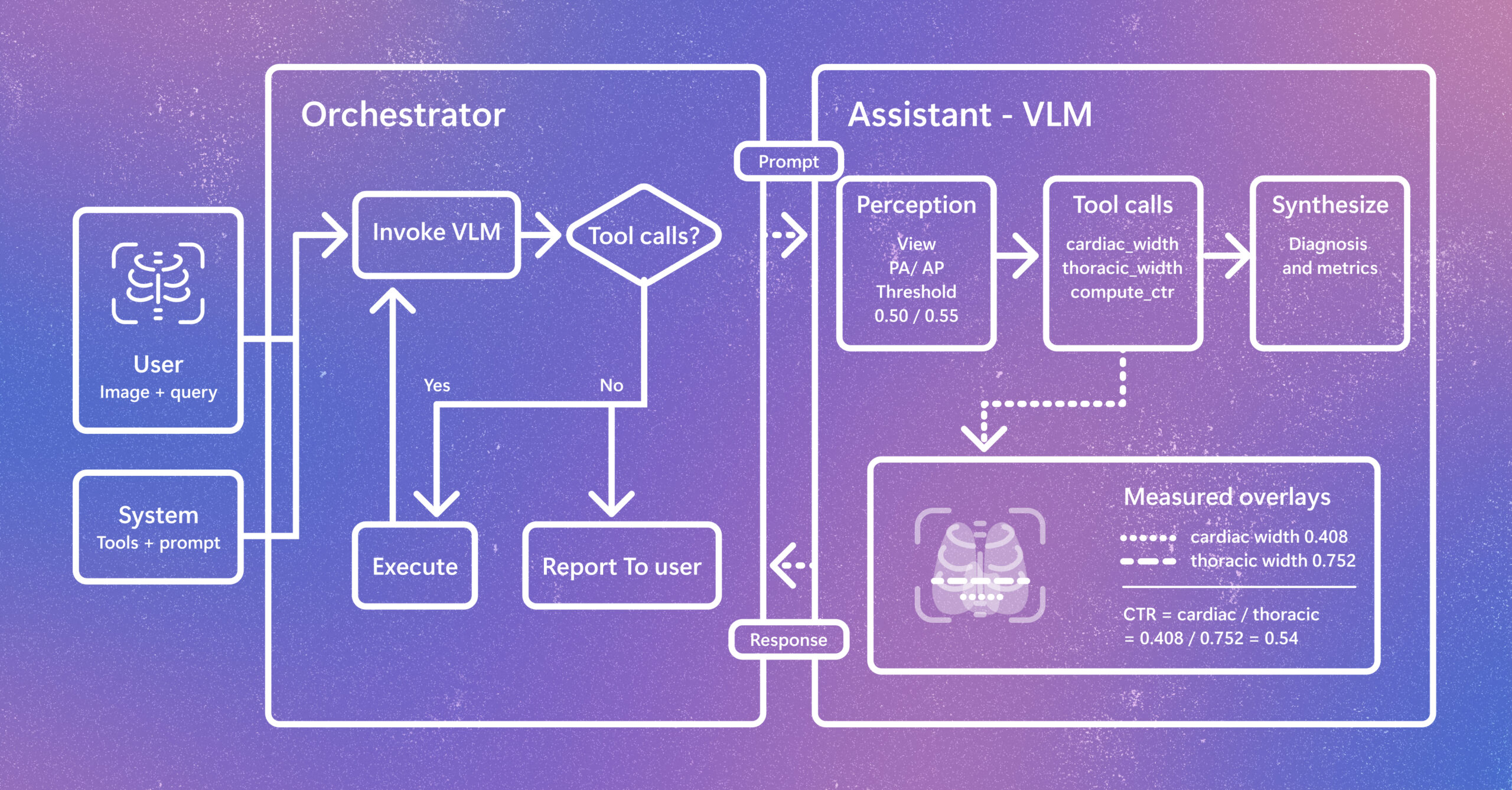
Radiology AI is evolving beyond report generation. CARE-X explores a unified approach that combines flexible reasoning, calibrated predictions, and measurement-based tools for chest X-ray interpretation. The post Introducing CARE-X: Towards Clinically Useful Radiology VLMs with Auxiliary Supervision, Reward-Aligned Learning, and Tool-Augmented Measurement appeared first on Microsoft Research.