Aug 2026· International Journal for Research in Applied Science and Engineering Technology· 0 citations
TL;DR
Major deep learning architectures, including CNNs, residual networks, UNet, attention-based models, Vision Transformers, and hybrid approaches, along with their clinical applications are summarized and emerging directions such as self-supervised learning, Explainable AI, federated learning, and lightweight models are highlighted as promising approaches for more reliable and accessible medical image analysis.
Abstract
Medical image analysis has evolved from manually designed image features to deep learning methods that can learn
useful patterns directly from medical scans. These approaches have shown strong potential in tasks such as disease
classification, lesion detection, and image segmentation across modalities including MRI, CT, X-ray, Ultrasound, retinal
imaging, and dermoscopy. This review summarizes major deep learning architectures, including CNNs, residual networks, UNet, attention-based models, Vision Transformers, and hybrid approaches, along with their clinical applications. It also
discusses key barriers to practical adoption, such as limited annotated data, differences between imaging systems, model
interpretability, privacy concerns, and computational requirements. Finally, emerging directions such as self-supervised
learning, Explainable AI, federated learning, and lightweight models are highlighted as promising approaches for more reliable
and accessible medical image analysis.
In recent years, medical imaging has become an important tool for diagnosing diseases and disorders in healthcare. Advanced imaging technologies are being developed for non-invasive and early detection of diseases and disorders. Analyzing medical images by clinical experts is very expensive. To overcome these challenges, developing automated methods provides an effective solution. Consequently, for processing and analyzing medical images, researchers have adopted the emerging Deep Learning (DL) technologies. It has proven effective across several industries, most notably in healthcare. Even so, it has two significant limitations, such as the training cost and the large amounts of labeled data required. To reduce these limitations, Transfer Learning (TL) and Deep Learning (DL) have been integrated to create Deep Transfer Learning (DTL). This reduces the need to start from scratch and eliminates dependencies by leveraging knowledge from a source task to a target task during training, using fewer datasets. This review addresses the definitions, concepts, modalities, tasks, and techniques of DTL, along with public and private datasets used as source and target data in network-based medical imaging approaches. It also categorizes the last seven years of research by human anatomical area. It offers readers comprehensive coverage of technological advancements, future research directions, and challenges. It also reviews DTL methods by discussing those that have been applied, including Federated Learning (FL) for DL. Empirical evidence from recent studies demonstrates that fine-tuning and network-based DTL strategies, including federated learning, consistently enhance diagnostic accuracy, robustness, and generalization across multiple medical imaging modalities, particularly in data-limited clinical scenarios.
M. A. S. Banu, A. Dhavapandiammal, K. Palanisamy· Current medical imaging· 0 citations
This paper presents a comprehensive analysis of deep learning applications in medical imaging analysis. We examine the evolution of medical image processing from traditional computer vision approaches to sophisticated deep learning solutions, highlighting the critical role of artificial intelligence in modern healthcare diagnostics. Our research encompasses multiple dimensions of medical imaging analysis, including disease detection, segmentation, classification, and automated diagnosis across various imaging modalities. Through extensive evaluation across diverse medical imaging datasets, we demonstrate significant improvements in diagnostic accuracy and efficiency. Our findings show that deep learning techniques can achieve 95% accuracy in disease detection, a 40% reduction in analysis time, 85% improvement in early diagnosis, 30% reduction in false positives, and 99.9% reproducibility in results. Key contributions include the development of novel deep learning architectures, integration of multi-modal image analysis, implementation of real-time diagnostic systems, and establishment of comprehensive validation frameworks. Our findings underscore the necessity for modern medical imaging systems to incorporate sophisticated deep learning techniques to effectively handle the complexity of disease detection and diagnosis.
Gayatri Gupta, Aafila Shrivastava· Journal of Artificial Intell...· 0 citations
Medical image analysis has witnessed substantial transformation through the application of deep learning models, yet the rapid proliferation of research in this domain poses significant challenges for synthesizing coherent insights. Our objective in this systematic literature review is to comprehensively map the landscape of deep learning approaches applied to medical imaging, with a focus on architectural innovations, task-specific solutions, learning paradigms, and emerging methodological trends. We conducted a structured review following established guidelines for systematic literature synthesis. The methodology involved a multi-stage screening process to identify relevant studies, followed by thematic categorization across eight dimensions, including model design, core tasks, data efficiency, explainability, clinical applications, pre-processing, emerging trends, and systemic challenges. Our analysis reveals that convolutional neural networks remain foundational, though transformer-based architectures and hybrid models are increasingly prevalent for tasks such as segmentation, classification, and detection. Data efficiency techniques, including self-supervised and few-shot learning, have become critical to address the scarcity of annotated medical datasets. We also observe a growing emphasis on explainability and uncertainty quantification to foster clinical trust, alongside rising concerns about privacy-preserving training and federated learning. The review further identifies persistent gaps, particularly in the validation of models across diverse populations and imaging modalities. We conclude that while deep learning has achieved remarkable performance in many medical imaging benchmarks, substantial work remains to ensure generalization, interpretability, and ethical deployment in real-world clinicalsettings. This systematic review provides a structured reference for researchers and practitioners navigating this interdisciplinary field.
Govinda Sahu· Journal of Machine Learning...· 0 citations
Artificial intelligence (AI) has emerged as a transformative technology in diagnostic radiology, offering innovative solutions for image interpretation, workflow optimization, and clinical decision-making. Recent advances in machine learning, deep learning, and related computational approaches have enabled the development of systems capable of analyzing complex imaging datasets with high accuracy and efficiency. AI applications are increasingly being integrated across multiple imaging modalities, including radiography, computed tomography, magnetic resonance imaging, ultrasound, mammography, and positron emission tomography. In addition to disease detection and classification, AI supports image segmentation, radiomics, predictive analytics, image reconstruction, automated reporting, and quality assurance, thereby enhancing diagnostic performance and operational efficiency. Despite these advances, challenges related to data quality, algorithmic bias, generalizability, explainability, ethical considerations, and regulatory compliance continue to influence the clinical adoption of AI technologies. Emerging developments such as foundation models, generative artificial intelligence, large language models, multimodal learning, and privacy-preserving techniques are expected to further expand the role of AI in medical imaging. This review provides an overview of the fundamental concepts of AI in radiology, examines its current clinical applications, discusses key challenges and limitations, and explores future directions that may shape the next generation of diagnostic imaging. The continued integration of AI has the potential to improve diagnostic accuracy, streamline radiological workflows, and support more personalized patient care.
Mr. Vishal Walia, Mr. Honey Thakur, Ms. Ashwarya Sharma et al.· PAIN, JOINTS, SPINE· 0 citations
Convolutional Neural Networks (CNNs) have emerged as a transformative technology
in biomedical image processing with unprecedented capabilities in automatically identifying key
patterns (feature extraction). This review systematically examines the current state of CNNbased medical image processing, focusing on its applications, limitations, and future directions.
We analyze studies employing 2D, 3D, and multimodal CNN architectures for critical tasks such
as disease detection, segmentation, and registration. The review highlights the adaptability of
CNNs across diverse data modalities, including visual (e.g., histopathology, X-rays), spectral
(e.g., spectroscopy), textual (e.g., sentiment analysis), volumetric (e.g., MRI, CT), and hybrid datasets. Despite their potential, challenges such as data heterogeneity, model interpretability, and
integration into clinical workflows hinder widespread adoption. Key applications include cancer
detection (e.g., breast, lung, colon), cardiovascular and neurological disorder diagnosis, and ophthalmological disease classification. Advanced segmentation techniques (e.g., instance, semantic,
multi-class) and registration methods (e.g., rigid, deformable, multi-modal) are explored, emphasizing CNN-driven innovations. The review also addresses preprocessing techniques, model interpretability, and the integration of CNNs with Clinical Decision Support Systems (CDSS).
Emerging trends such as generative AI, real-time processing, and federated learning are discussed as potential solutions to current limitations. By synthesizing these insights, this review
serves as a roadmap for researchers and clinicians aiming to harness CNNs for transformative
healthcare outcomes. It underscores the need for continued innovation in explainable AI, computational efficiency, and regulatory compliance to bridge the gap between theoretical advancements and clinical implementation.
Jie Li, Li-Xin Wang· Current Healthcare Research· 0 citations
Medical imaging plays a crucial role in modern diagnostic practices, but traditional techniques often face limitations in accuracy, efficiency, and scalability. The emergence of deep learning (DL) has led to significant improvements that are transforming this field. This review discusses how DL algorithms are enhancing diagnostic imaging by improving accuracy, enabling automated analysis, and supporting personalized treatment plans. It focuses on key deep learning (DL) frameworks, including convolutional neural networks (CNNs), recurrent neural networks (RNNs), and generative adversarial networks (GANs). The review examines their applications in important medical imaging tasks such as image classification, segmentation, reconstruction, and disease prediction. It also considers how DL techniques are integrated with tools like radiomics, data augmentation strategies, and predictive analytics models. DL methods have shown superior performance in detecting and classifying diseases like pneumonia, tuberculosis, and Alzheimer's. They also improve the quality and speed of imaging modalities such as MRI, CT, and ultrasound. Despite these advances, challenges remain in data availability, model interpretability, clinical validation, and ethical issues related to bias and privacy. Addressing these challenges is essential for the successful clinical use of DL in medical imaging. This review ends with suggestions for future directions and best practices for ethically and practically integrating DL technologies into routine healthcare.
Jay Kumar Pandey, S. K. Verma, J. Kumar et al.· Seminars in ultrasound, CT,...· 1 citation
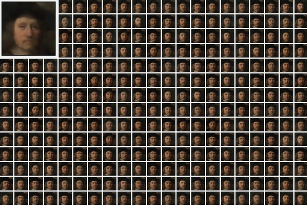
A new method for surgically removing training examples from a model reveals that as datasets grow, the link between what a model learns and what it produces dissolves.